Architecture of the Kidney in Chronic Bright's Disease
- SIGNED cloth binding
- New York: Paul B. Hoeber Inc., 1939
New York: Paul B. Hoeber Inc., 1939. First edition.
SCARCE ASSOCIATION COPY--BEAUTIFULLY ILLUSTRATED STUDY OF CHRONIC KIDNEY DISEASE BY DETAILED NEPHRON MICRODISSECTIONS.
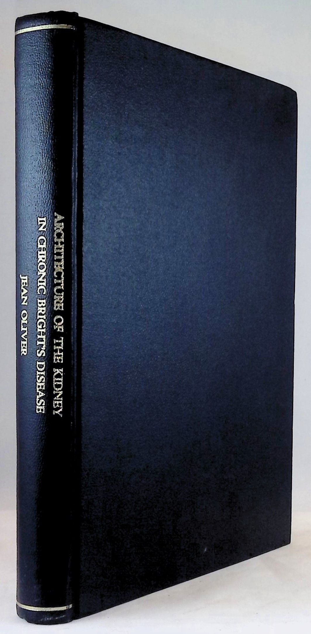
11 1/2 inches tall hardcover, black pebbled cloth binding, gilt title to spine, signed on front free endpaper, "Benjamin H. Landing/ (Gift of Dr. George Fetterman)". i-xiv, 257 pp, 112 illustrations including 5 color and 39 aquatone plates (a lithographic process for printing by offset from a metal plate coated with photosensitized gelatin). Near-fine.
JEAN REDMAN OLIVER (1889-1976) earned his AB and MD from Stanford University, where he remained on faculty from 1914 to 1929, interrupted in 1916-1919 by studies at the Rockefeller Institute. From 1929 to 1950, he was Head of the Department of Pathology at Long Island College of Medicine, then moved to the State University of New York Medical College where he remained until retiring as professor emeritus 1955. This was followed by 17 years of continued productivity, as he established his laboratory at the Overlook Hospital in Summit, New Jersey, and was joined by his brilliant assistant, Muriel MacDowell, who mastered the microdissection techniques. They outgrew their quarters at Overlook Hospital, and became investigators of CIBA Pahrmaceuticals in Summit, supported by NIH grants. Oliver was a highly innovative pathologist who played a critical role in the development of nephrology in the 20th century by appreciating the inportance of nephron heterogeneity in kidney disease, and the correlation of nephron structure with function. The latter became possible by his collaboration with renal physiologists (A.N. Richards, A.M. Walker, and C.W. Gottschalk) who developed single nephron micropuncture techniques.
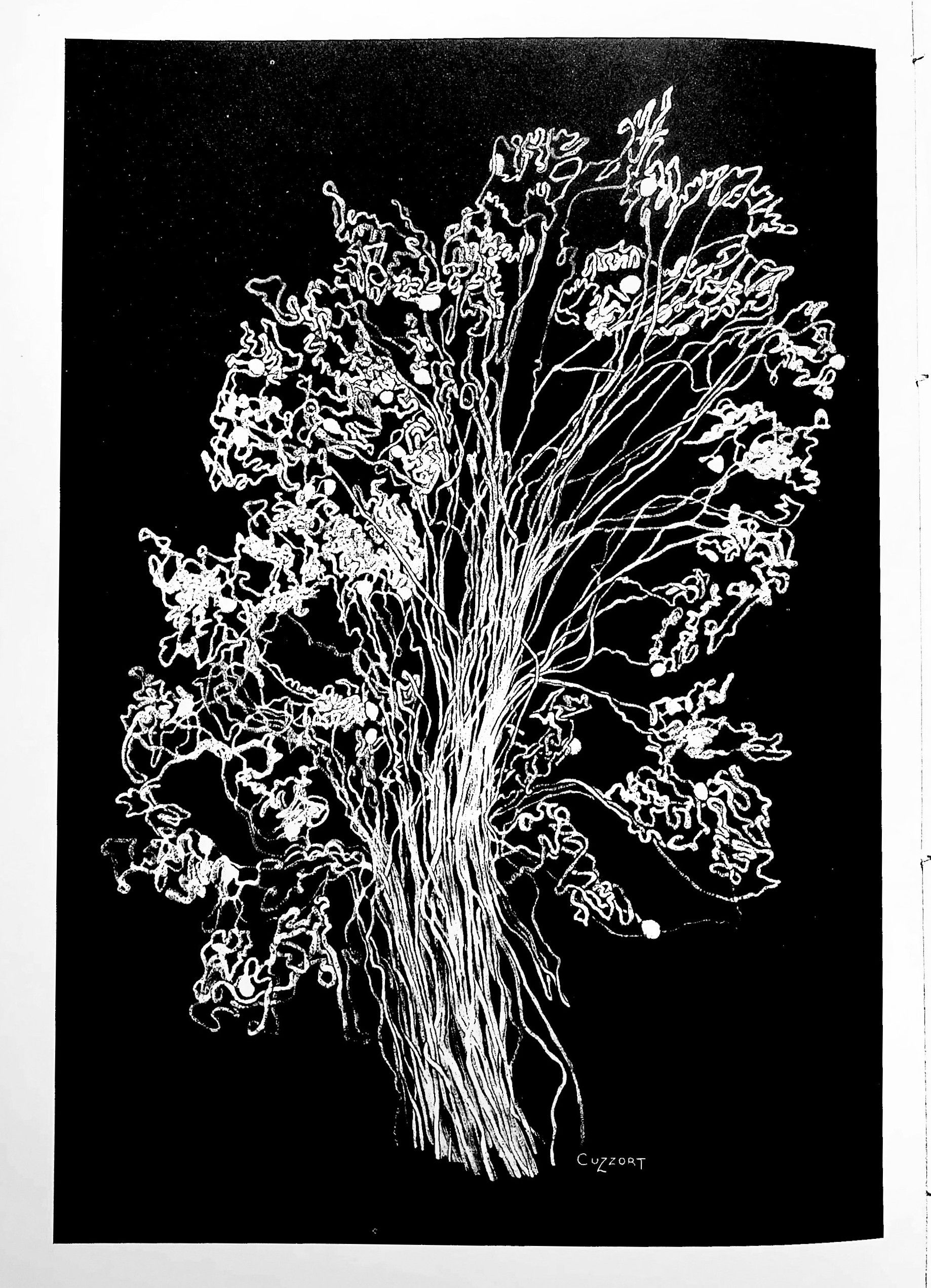
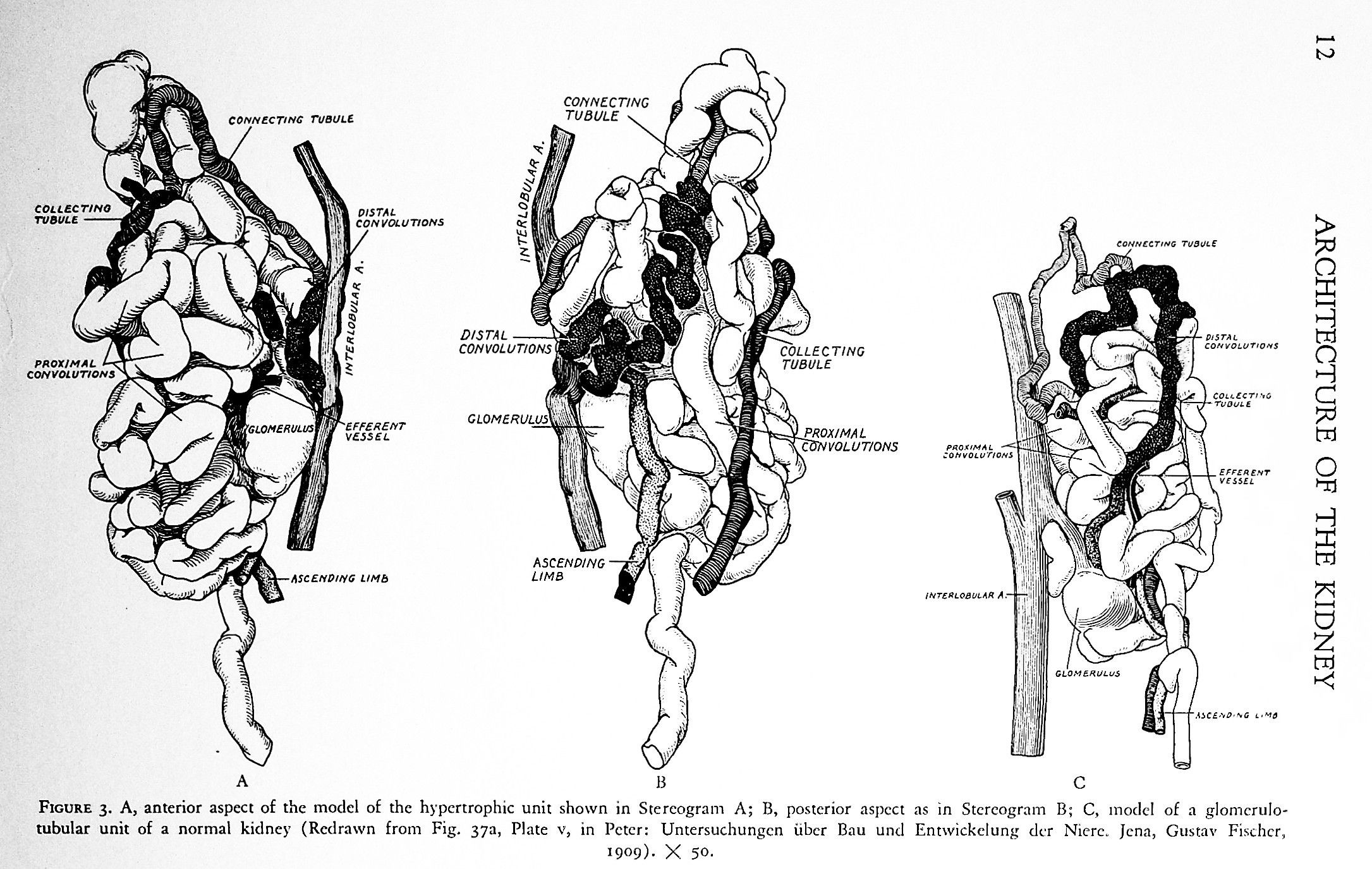
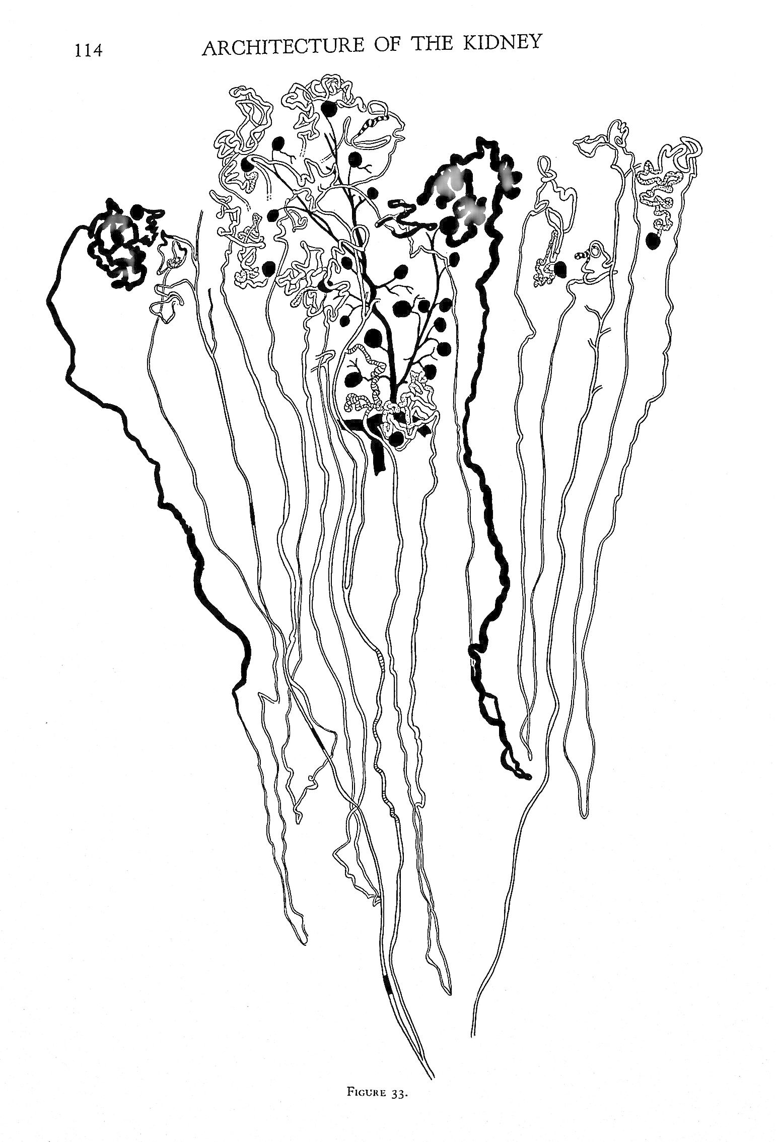
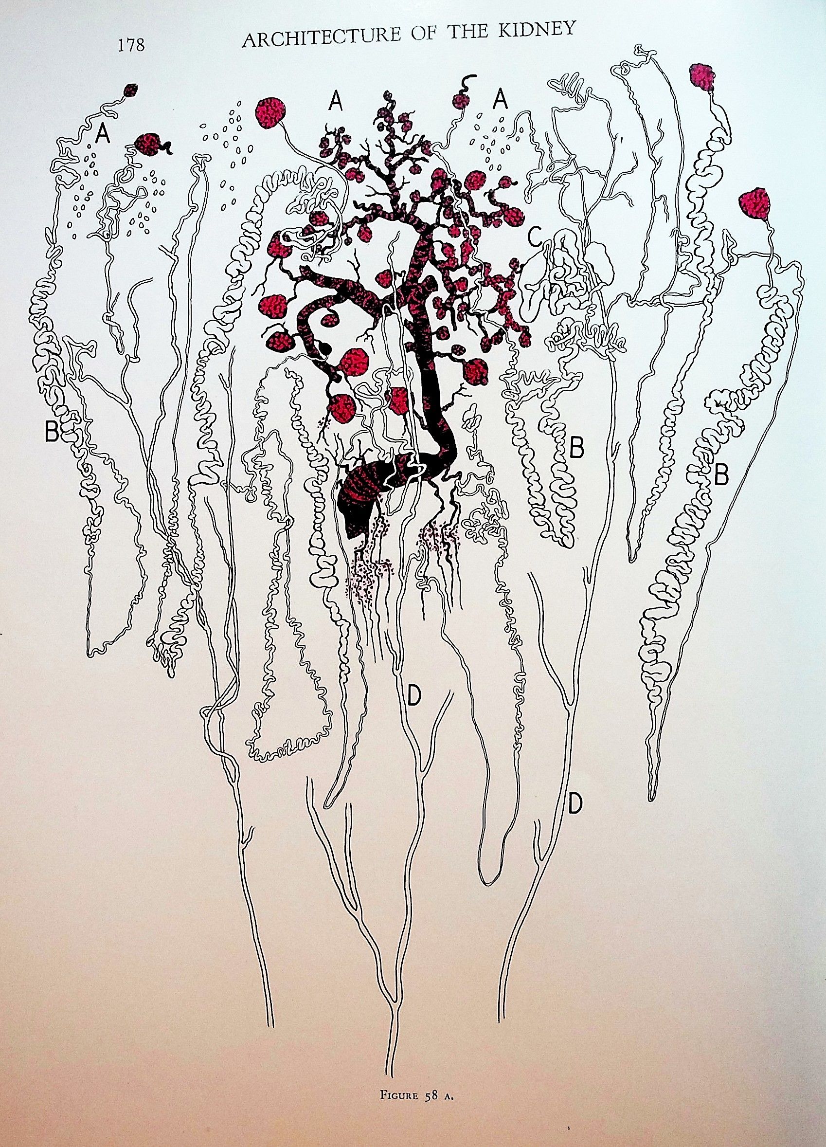
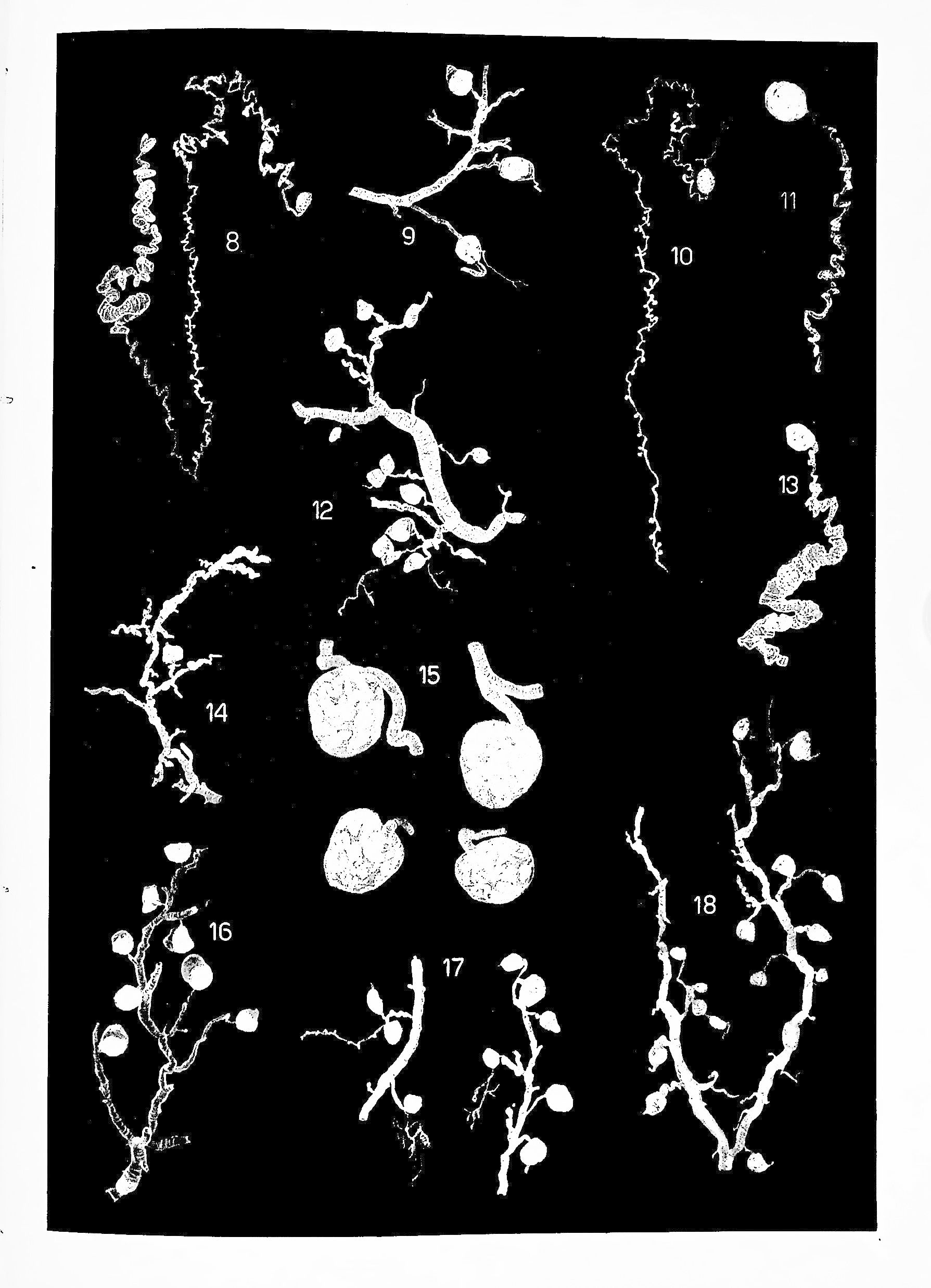
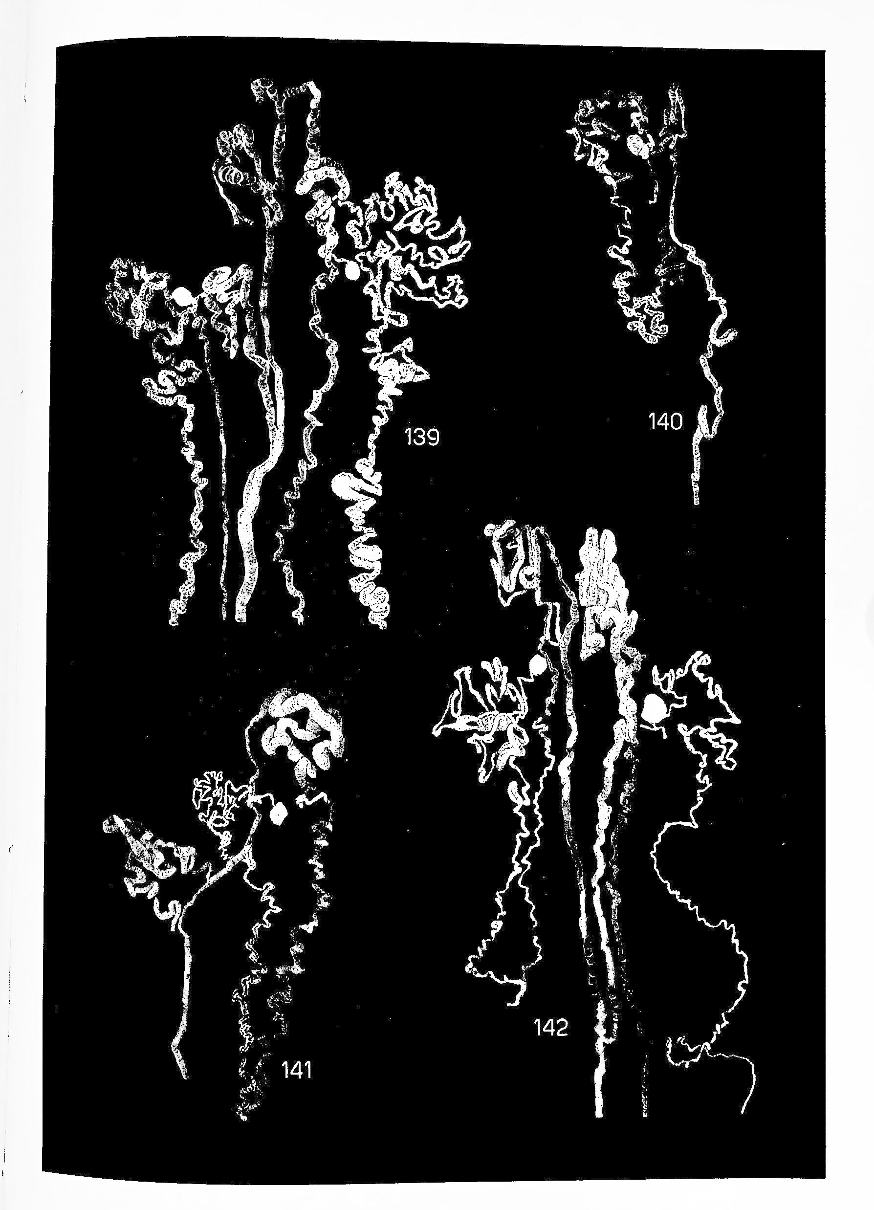
CITED BY S.E. BRADLEY in JEAN REDMAN OLIVER IN CONTEXT, Kidney International, Vol. 5 (1974) pp77-95: "As Oliver pointed out in his Hektoen Lecture in 1934, it was the application of the maceration and microdissection technique and the Born wax plate reconstruction that permitted Hiiber in the United States and Peter in Germany to describe the normal nephron in detail. ... It seemed reasonable to believe they would add as much to an understanding of pathology Oliver and his associates—among whom a remarkably skilled and artistically gifted colleague, Muriel MacDowell, became increasingly more prominent--perfected their technique carefully, innovatively and productively. The dissected nephrons were now stained affording cellular detail not only to be seen but registered objectively in ingenious mountings of photomicrographs, thus extending its applicability. A series of successes was achieved with the improved methodology and recorded in a number of papers that Oliver summarized in his monograph of 1939, The Architecture of the Kidney in Chronic Bright's Disease. The material presented consists of illustrations of more than 300 nephrons of the thousands dissected from contracted kidneys of approximately 40 patients dying in uremia or from other causes, taken for the most part from the series previously studied with Addis and described in clinical and pathological detail in The Renal Lesion in Chronic Bright's Disease. [Descriptions of individual microdissected kidneys in Architecture of the Kidneys reference clinical details in case numbers in The Renal Lesion] Although an amazingly varied array of nephron and vascular deformities was examined, a striking similarity was evident in the architecture of all forms of contracted kidneys, 'no matter what combination of initial processes has produced the ultimate deformity.' The figures showing microdissections of a "lobule" from a normal adult kidney (Fig. 2) and of clusters of defective nephrons in their entirety from 23 contracted kidneys to show the pattern of renal architecture in each disease process are stunning evidence of a technical tour de force."
PROVINANCE: BENJAMIN HARRISON LANDING (1920-2000), pioneering pediatric pathologist, attended Harvard Medical School, obtaining his M.D. cum laude in 1945. He proceeded immediately into pediatric pathology, serving his internship in Sidney Farber's department at the Children's Hospital in Boston. In 1948 he returned to complete his residency training at Boston Children's Hospital and at the Free Hospital for Women and Boston Lying-in Hospital. In 1953 Landing was selected to develop the Anatomical Pathology Department at Cincinnati Children's Hospital. By the time he moved to Children's Hospital, Los Angeles, in 1961, he had published some 80 peer-reviewed papers.
GEORGE H. FETTERMAN (1908-1988) graduated from the University of Pittsburgh School of Medicine in 1930 and interned at Mercy Hospital of Pittsburgh. His training in pathology included a year of residency at Mercy Hospital and two years at the University of Toronto. Between 1934 and 1950 he was Director of Laboratories, first at Pittsburgh City Home & Hospitals and then at St. Margaret Memorial Hospital, in Pittsburg. In 1937 he was elected a Fellow of the American Society of Clinical Pathologists. Much of Doctor Fetterman's investigative work involved microdissection of the kidney. He was a master of that arcane art and studied normal morphogenesis as well as several renal diseases. Many of his contributions remain central to our understanding of those conditions
Details
Title
Architecture of the Kidney in Chronic Bright's Disease
Author
Oliver, Jean
Binding
cloth binding
Condition
Unknown
Publisher
Paul B. Hoeber Inc.: New York
Date
1939
Edition
First edition